Sexing Tarantulas Using Molts
A tarantula once it has reached a certain size can be reliably sexed using its cast-off exoskeleton; the molt or exuviae. For this how to I used a molt of a juvenile Brachypelma smithi that molted the 25th of June, 2020.
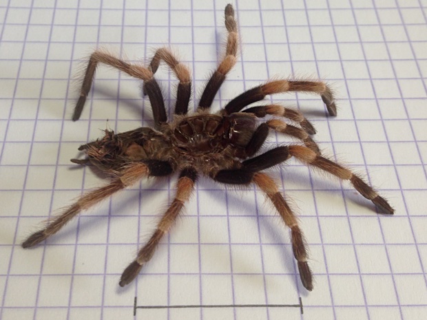
In the above photo each square on the paper is 5mm by 5mm. The black line near the bottom is 25mm or about an inch. This should give you an idea of the size of the molt used. For smaller molts a microscope might be required.
In order to determine the sex we look inside the abdomen for the presence of the female's spermathecae. The abdomen is the "hind" part of the tarantula. Because this part can be twisted and stuck together we need to manipulate it. This goes easiest if the molt is moist and soft. In order to achieve this I put the molt in a dish with some water and dropped some water on the abdomen as well. I let it soak for about 10 minutes.
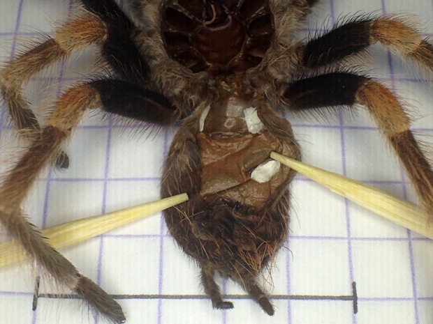
The area of importance is between the first pair of book lungs; the large white spots in the above photo. Try to smooth this area as much as possible. I use toothpicks to carefully manipulate the molt. Moreover, try to wick away water as much as possible using the side of a piece of toilet paper or a tissue.
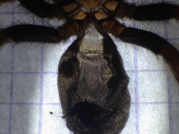
Next, place the molt carefully on a piece of paper and use a flashlight to shine light underneath the paper. Position the flashlight in such a way that the area of interest receives most of the light. Make sure that the area of interest has been dried using the wicking method as mentioned above.
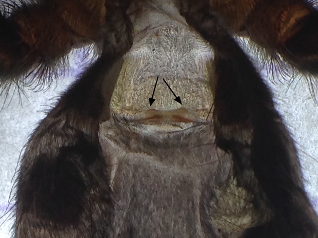
In the above photo you can clearly see the spermathecae making this specimen a juvenile female Brachypelma smithi. I used an iPhone 5 with a macro lens and a LED ring light for the above two photos. The latter two I bought for less than 4 euro as a Multi lens set for smartphone.
Note that the last photo is an 1:1 crop of the previous one. I recommend that if you ask for help with sexing, for example on Facebook, that you make a copy of the photo and then crop (cut out) the area of interest. With 1:1 I mean that each pixel in the above photo corresponds with one pixel in the original, so no scaling took place. I recommend cropping because Facebook drastically reduces the resolution of uploaded photos. So while the area of interest might apear sharp when you zoom in on your phone or camera it will be very grainy when zoomed in on Facebook.
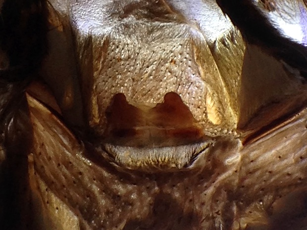
The actual shape of spermathecae differ from species to species hence it is a good idea to compare what you see with photos of spermathecae of the same species. For example, in the above photo you can see the spermathecae of an Aphonopelma seemanni. This exuviae is from a (sub) adult female; the spermathecae could easily be seen with the naked eye.